APD based technology with Depth-of-Interaction capability
What is the benefit of Time-of-Flight?
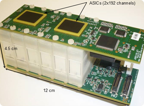
Our Avalanche Photodiode (APD) based technology for PET was developed by a R&D Consortium including PETsys S.A., with a focus on research and development. The R&D Consortium licensed this technology to PETsys SA. The APD-based photon detectors use matrices of LYSO scintillation crystals coupled to APD arrays. The APD signals are readout and sampled by a dedicated ASIC with 192 channels.

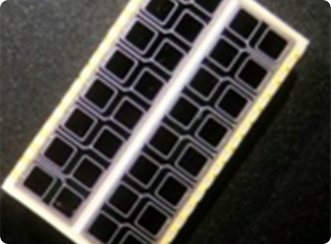
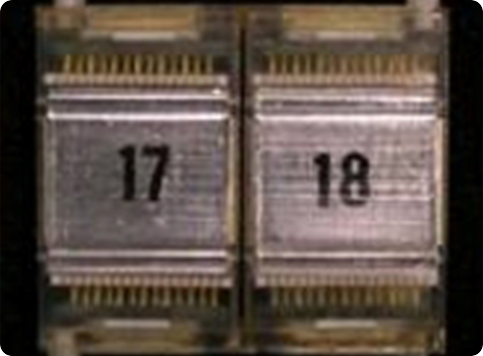
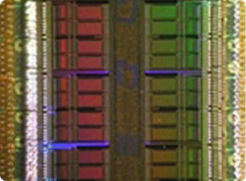
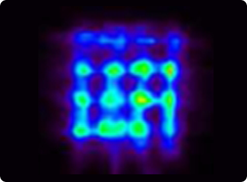
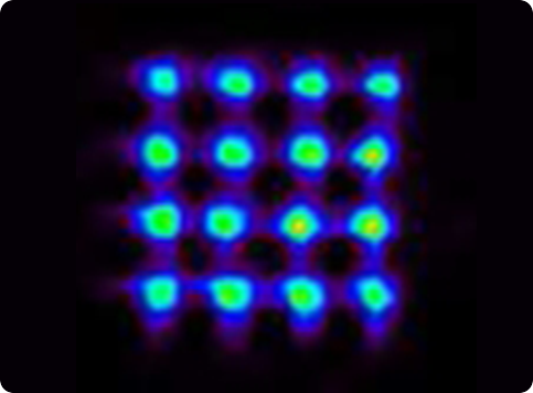
Several crystal-APD units and the corresponding frontend electronics are assembled in detector modules. Our APD-based detector module has 384 LYSO 2x2x20 mm crystals and covers a detection area of ~4x8 cm. One of the advantages the APDs is the capability of performing the double readout of the scintillation crystals, which allows to calculate the DoI of the detected photons. Associated to the fine detector pitch (2x2 mm) and the one-to-one coupling between crystal and APD pixels, the DoI capability permits an excellent spatial resolution of the order of 1.3 mm over the whole Filed-of-View.
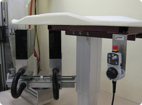
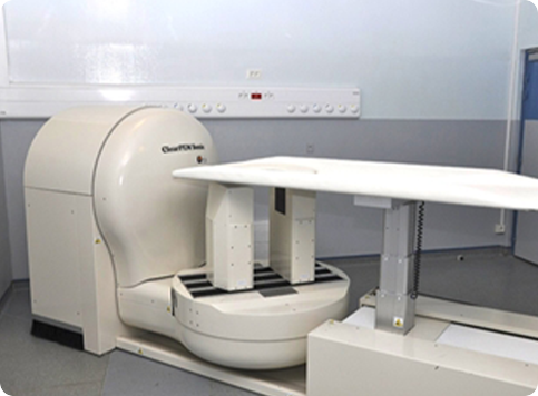
PETsys has developed two PET prototypes based on APD-based detector modules which target breast cancer applications (Clear PEM System).
These systems comprise 12288 APD's and 6144 scintillation crystals, organized into two opposing detector plates. One of these is located at ICNAS in Coimbra (PT) - (Fig. 5).
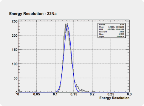
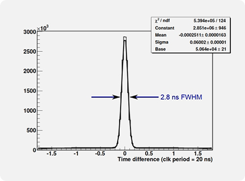
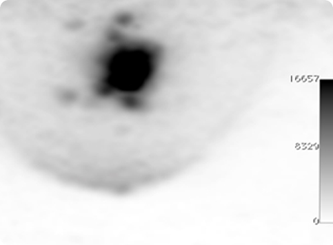
The main spectroscopic characteristics of the Clear-PEM system are energy resolution for 511 keV photons of of 13.4%, coincidence time resolution of 2.8 ns (FWHM) and DoI resolution of 2.8 mm (FWHM).
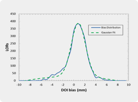
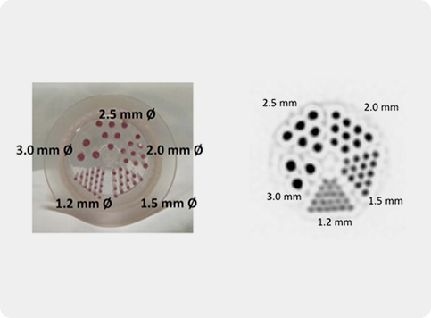
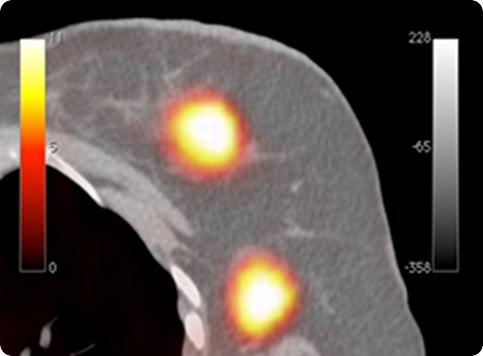
Clear-PEM double readout scheme allows calculation of photon Depth of Interaction (DOI) and compensates blurring due to the parallax effect in PET resulting in an isotropic image resolution of 1.3 mm FWHM.
PETsys and the R&D Consortium were invited by CERN to join the Clear-PEM Sonic project combining PET with Ultrasounds, in the framework of the Crystal Clear Collaboration for detection of early stage breast cancer. We have developed the first integrated PEM-Ultrasound system with Elastography capabilities, the Clear-PEM Sonic prototype, along with SuperSonic Imagine. This system is now being tested at Ospedale San Gerardo in Italy after being used in a series of clinical tests at Hospital Nord in Marseille.
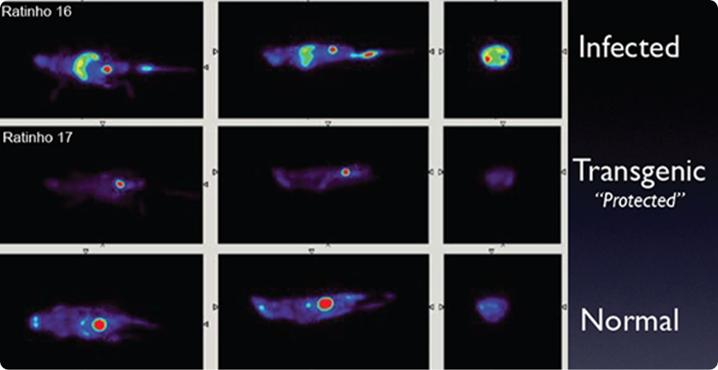
Due to its very good spatial resolution and accessibility of the Field-of-View, the Clear-PEM system can also be used for small animal imaging.